Cutaneous and Mucosal Leishmaniasis

Cutaneous leishmaniasis (CL) and mucosal/mucocutaneous leishmaniasis (ML) are infectious diseases that affect the skin and mucous membranes.

They are caused by protozoa of the genus Leishmania and are transmitted to animals and humans by vectors of the Psychodidae family. Their distribution is worldwide, and they are endemic in 89 countries. In 2021, 51 countries reported nearly 222.000 new autochthonous cases to the World Health Organization.

Among the nine countries in the world with the highest number of cases of cutaneous leishmaniasis, three are in the Americas: Brazil, Colombia, and Peru. In the Region of the Americas, an annual average of 52.645 cases of cutaneous and mucosal/mucocutaneous leishmaniasis have been recorded in the last 20 years, with a gradual downward trend in cases since 2005. In 2021, 37.786 cases of cutaneous leishmaniasis were reported, 22.5% of them in border areas.

Cutaneous leishmaniasis has been recorded in 21 of the Region's countries, and is endemic in 19 (Argentina, Belize, Bolivia, Brazil, Colombia, Costa Rica, Ecuador, El Salvador, Guatemala, French Guiana, Guyana, Honduras, Nicaragua, Mexico, Panama, Paraguay, Peru, Suriname, and Venezuela). It should be noted that French Guiana reports its data directly to France. 

Read Leishmaniases: Epidemiological Report of the Americas

Detailed information


CL and ML are caused by different species of protozoa of the genus Leishmania, comprising approximately 22 species that are pathogenic to humans. In the Americas, human infection is caused by 15 species, which are grouped into the subgenera Leishmania and Viannia

In the Region of the Americas, the three most important species of the subgenus Leishmania are: L. (L.) mexicana, L. (L.) amazonensis, and L. (L.) venezuelensis. The subgenus Viannia has four main species: L. (V.) braziliensis, L. (V.) panamensis, L. (V.) peruviana, and L. (V.) guyanensis. The species are morphologically indistinguishable, but can be differentiated by isoenzyme analysis, molecular methods, or by monoclonal antibodies and PCR testing.

Leishmania infections that cause CL and ML have been described in several species of wild and synanthropic animals, and, in certain specific transmission cycles, in domestic animals. Some species of rodents, marsupials, and edentates have been identified as natural hosts and potential sylvatic reservoirs.

In the Americas, leishmaniasis is transmitted predominantly by the bite of sandflies of the genus Lutzomyia, according to the classification by Lewis and revised by Young. However, Galati's classification recognized 23 genera, with around 54 species involved in transmission.

The incubation period in humans is two to three months on average, but it can be shorter (two weeks) or longer (two years).

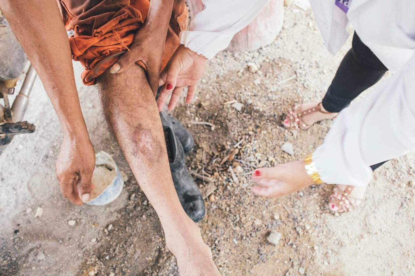
People with CL may have one or more skin lesions, and the ulcers can change in size and appearance over time. When the phlebotomine sandfly vector bites a person, it causes a macule, which evolves into a papule, and then into a rounded, painless nodule that progressively increases in size and ulcerates. Initially the ulcers are covered by a scab, and when detached, the typical ulcer has a clean background, pink color, and granulous tissue; it is round, has regular, raised margins, is painless, and has an indurated base. Ulcers can sometimes become secondarily infected with other microbial agents.

When the disease involves the pinna, mutilation may occur. This type of lesion was initially described as the "chiclero ulcer" and is very frequent in the Mexico's Yucatan Peninsula.

In mucosal lesions, the initial and most frequently affected site is the mucosa of the nasal septum, which can progress to perforation. The process can extend to the palate and pharynx; the uvula is infiltrated, hypertrophied and then amputated. When it attacks the nose, it can cause obstruction, bleeding, runny nose, and the appearance of scabs and wounds. Involvement of the larynx and pharynx can cause pain, hoarseness, dysphonia, and dysphagia.

Clinical

Consideration should be given to people from endemic areas who have signs and symptoms suggestive of CL and ML.

Laboratory

Parasitological tests are used to view the parasite (direct parasitology, culture, and PCR). In the case of skin lesions, the samples can be smears, aspirates, or biopsies obtained from the lesion, depending on the examination to be performed. For the diagnosis of mucosal or mucocutaneous leishmaniasis, the diagnosis is clinical, complemented by specific parasitological, histological, and/or immunological tests.

No vaccines are available to prevent infection.

Human

Recommended individual or collective protection measures for people are aimed at reducing contact with vectors. In particular, it is important to avoid outdoor activities from dusk to dawn; use mosquito nets; use protective clothing and insect repellents; and build dwellings at least 400 meters from forest boundaries or areas with dense vegetation.

Vectors

Carry out environmental management (cleaning yards and grounds) to change the environmental conditions that provide breeding sites for immature forms of the vector. In the rural domestic environment, keep animal shelters secluded and maintain a consistent high level of cleanliness.

In 2022, PAHO published new Guidelines for the Treatment of Leishmaniasis in the Region of the Americas, detailing treatment regimens and indication criteria for leishmaniasis for the Region, consistent with WHO standards for guideline development. Some of the recommendations presented here may differ from the specific recommendations for other continents, given the Region's distinct epidemiological and biological characteristics, including the different circulating species of Leishmania, transmission cycles, and treatments.

Treatment may be local or systemic, depending on the clinical form of the disease and the local use criteria. Recommendations are for pediatric and adult patients and, when available, based on the species of Leishmania presumptively involved.

Read more about Recommendations for the treatment of cutaneous and mucosal/mucocutaneous leishmaniasis, as well as therapeutic regimens, routes of administration, and dosages.

More information