SUBMENU
Lymphatic Filariasis due to Wuchereria bancrofti is the only type of lymphatic filariasis present in the Americas.
The infection is transmitted by infected mosquitoes and is associated with acute and chronic symptoms that can lead to disfigurement and consequently to social exclusion and stigma.
Measures are being taken to eliminate lymphatic filariasis as a public health problem from the Americas. The main pillar of the strategy is the massive annual distribution of two anthelmintic medications, diethylcarbamazine, and albendazole, to all people living in endemic areas for five years, or three medications, the two previous plus ivermectin, for two years.
- Lymphatic filariasis, often known as elephantiasis, is a human infection that is caused by the transmission of parasites called filarias through mosquitoes, including those of the genus Culex, which is widespread in urban and semi-urban areas.
- Mosquitoes become infected with microfilariae by ingesting blood when an infected carrier is bitten. The microfilariae mature in the mosquito and become infectious larvae. When infected mosquitoes bite people, mature parasite larvae settle on the skin, from where they can penetrate the body.
- Lymphatic filariasis adopts asymptomatic, acute and chronic forms. Most infections are asymptomatic and have no external signs. Despite this they damage the lymphatic system and the kidneys and alter the immune system. The painful and very disfiguring manifestations of the disease, lymphedema, elephantiasis and scrotal inflammation appear later and can cause rejection and social stigma, with the consequent loss of self-esteem and decreased job opportunities for those affected, and therefore, affected their economic and social situation.
- The disease can be eliminated in the Americas with the simultaneous massive administration of two anthelmintic drugs, diethylcarbamazine and albendazole (DA) to all people living in endemic areas for 5 years or three medications, the previous two plus ivermectin (IDA), for 2 years.
- In places where the disease is endemic, the use of mosquito nets is recommended in the windows and doors of homes and on beds, the elimination of mosquito breeding sites and the application of insecticides in open latrines.
Lymphatic filariasis (LF) is a parasitic infection caused by worms (nematodes) which can lead to changes in the lymphatic system and trigger chronic lymphedema, abnormal enlargement of parts of the body (elephantiasis), pain, severe disability, stigma, and social exclusion.
In the Americas, Wuchereria bancrofti is the only worm species transmitted by the Culex mosquito (mainly of the species quinquefasciatus) which are the most common vectors. Adult worms in the human host are whitish or pinkish, thin (1.5-2.5mm), and can measure up to 1 meter in length.
Only four countries are endemic to lymphatic filariasis in the American continent:
- Dominican Republic
- Guyana
- Haiti
It is estimated that up to 12.6 million individuals are at risk of infection. Costa Rica, Suriname and Trinidad and Tobago were eliminated from the list of endemic countries by PAHO/WHO in 2011.
In order to complete its life cycle, W. bancrofti requires a definitive host (the man, as there is no significant animal reservoir) and a vector (Culex quinquefasciatus is the main vector responsible for transmission of lymphatic filariasis in the Americas). Lymphatic filariasis is transmitted when the third-stage larvae (L3) of the worm are deposited on the skin by an infected mosquito vector at the time that the mosquito is feeding.
The larvae then penetrate the skin at the bite, migrate to the lymphatic system and in approximately a year mature into adult male and female worms. Adult females release larvae (called microfilariae) that eventually migrate into the bloodstream and can reach a maximum concentration between 10pm and 2am (nocturnal periodicity). Ingestion of circulating microfilariae by another mosquito vector and their development into L3 larvae closes the life cycle of the parasite and sustains transmission in endemic areas.
Lymphatic filariasis is characterized by a chronic course, which is aggravated by highly-symptomatic acute episodes. The prepatent period (interval between the entry of the L3 larvae and the appearance of detectable microfilaraemia) can last several months. The presence of microfilariae in the blood may in fact be asymptomatic, while clinical manifestations, if present, can arise from a few months to a year after infection.
Clinical manifestations are usually classified into acute and chronic form:
Acute phase
- Recurrent attacks of fever with painful inflammation and swelling of the lymph nodes and ducts.
- Involvement of the genital tract is common
- Orquiepididimitis (inflammation of the testis and the epididymus) and funiculitis (inflammation of the spermatic cord) are typical findings in male patients.
- The affected lymph ducts appear dilated and with thickened walls.
These "acute attacks" last a few days and can be attributed to a combination of factors: presence of living adult worms, death of worms possibly complicated by bacterial infection, the immune system response to filarial or microfilarial antigens, etc.
Chronic phase
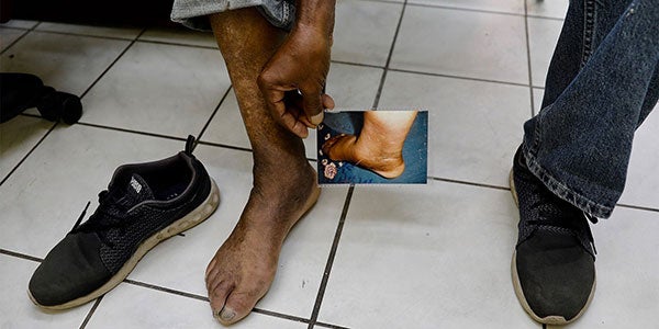
- Repeated episodes of inflammation that can lead to a progressive obstruction of the lymphatic ducts with accumulation of fluid in the interstitial tissues, resulting in lymphedema, a condition that most often affects the arms and the legs.
- The increasing fibrosis and sclerosis of the subcutaneous tissues can accelerate the progression of lymphedema to elephantiasis (thickening and hardening of skin fold in nodule formation and epidermal pigmentation changes), which is associated with excessively enlarged extremities and disability.
- In male patients, the obstruction of the spermatic lymphatic duct leads to accumulation of fluid in the scrotal sac (hydrocele).
- Stigma and social exclusion are frequent among such patients.
A suggestive clinical picture of the disease and a high eosinophil count may help the diagnosis. The standard technique used is the direct identification of microfilariae in thick blood smears. The blood sample must be collected near the time of maximum concentration of microfilariae in order to increase the sensitivity of the test.
There is the availability of FTS immunochromatographic tests (Filariasis Test Strips), which detect filarial antigens in blood collected after a finger prick, these are independent of the concentration of microfilariae and prove to be sensitive and specific. The ICT test can be used for mapping, monitoring, and evaluation of the transmission of lymphatic filariasis through community-level studies.
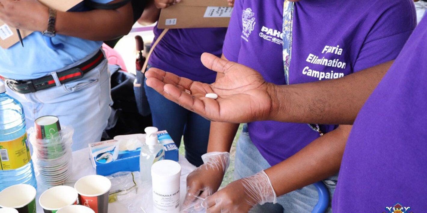
- Preventive treatment is based on combinations of 2 or 3 medications. The combination of 3 medications consists of ivermectin 150 μg / kg, diethylcarbamazine (DEC) 6 mg/kg and albendazole 400 mg in a single administration. The two-drug includes only diethylcarbamazine (DEC) and albendazole (ALB).
- Both combinations of medications are administered once a year. The triple administration reduces much more and for a longer time, the number of microifilariae in blood. This combination of drugs is associated with mild and temporary adverse reactions, but it is generally considered a very safe treatment.
- The significant effects of these two combinations of medicaments on the concentration of microfilariae in blood (or microfilaremia) are the reason why the strategy recommended by the WHO for the elimination of lymphatic filariasis is the massive annual distribution of one of these two combinations to all people living in an area where lymphatic filariasis is transmitted (microfilaremia is equal to or greater than 1%). When the triple combination is used, two annual rounds are required to interrupt transmission, while if double is used, at least five rounds of treatment are required annually.
- The interruption of the transmission can be accelerated with the additional application of mosquito vector control measures.
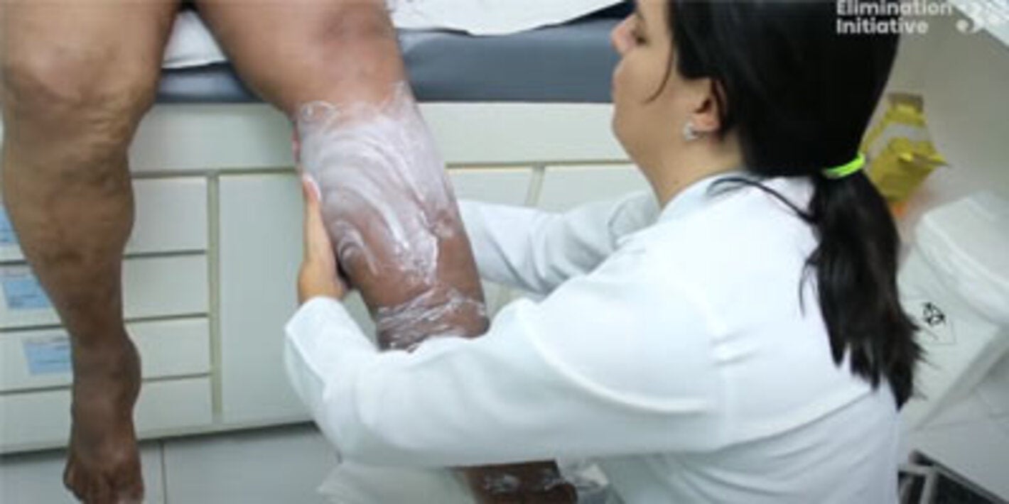
- In individuals who have already developed chronic manifestations such as lymphedema and elephantiasis, the management of the affected limbs is necessary to avoid further deterioration of health. This includes elevation, limb lavage, and treatment of skin infections. These measures can usually be performed by the patients themselves. Surgery is necessary in cases of hydrocele.
- In 1997 the World Health Assembly passed Resolution WHA50.29 on the elimination of lymphatic filariasis as a public health problem.
- In September 2016, through Resolution CD55.R9, the PAHO Directing Council approved the Action Plan for the Elimination of Neglected Infectious Diseases and Post-Elimination Measures 2016-2022, one of whose goals is the elimination of lymphatic filariasis in the Americas by 2022.
- In May 2013, the World Health Assembly adopted the Resolution WHA 66.12, and reaffirmed the goals of 2020 for 17 neglected tropical diseases, including Lymphatic Filariasis, by 2020.
- PAHO / WHO collaborates with endemic countries to obtain donations of drugs, diagnostic tests, and other supplies necessary to interrupt and eliminate transmission. PAHO also supports countries in the design and implementation of baseline epidemiological and impact assessment studies. Also, contributes to the planning, implementation, and evaluation of mass drug administration campaigns. PAHO / WHO coordinates these actions through the Regional Program of Neglected Infectious Diseases and supports endemic countries to obtain validation of the elimination of lymphatic filariasis as a public health problem.


